
非洲猪瘟病毒在掩埋野猪尸体中的存活情况(上)
来源:猪译馆 2020-06-12 14:36:14| 查看:次
译者的话:
非洲猪瘟病毒在掩埋野猪尸体中的存活情况(上)
African Swine Fever Virus Survival in Buried Wild Boar Carcasses – Part 1
作者Authors:
Claire Guinat, DVM, MSc, MRCVS
Andrey Gogin, DVM, PhD 欧洲食品安全局 European Food Safety Authority
Sandra Blome, DVM, Dr med vet
Guenther Keil, DipBiol, DrRerNat
Reiko Pollin, DipBiol, DrRerNat 里德里希·吕弗勒研究所 Friedrich-Loeffler Institut
Dirk U. Pfeiffer, PhD 英国皇家兽医学院兽医流行病学经济学和公共卫生研究组 Royal Veterinary College, Veterinary Epidemiology, Economics and Public Health Group
Linda Dixon, BSc, PhD, 英国皮尔布莱特研究院 The Pirbright Institute
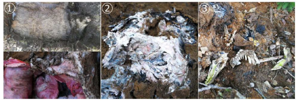
自从1957年非洲猪瘟(ASF)首次在欧洲野猪群中爆发以来,人们一直在讨论死于该病的动物尸体中病毒的存活问题。已知非洲猪瘟病毒(ASFV)在环境中非常稳定。因此,受感染野猪的尸体可以作为ASFV的主要宿主,从而有助于在野猪群中局部维持和传播该疾病。为了尽量减少这一风险,在ASF疫区清除野猪尸体被认为是有效控制疾病的关键。如果野猪尸体无法清除,通常会就地掩埋处理,以避免野猪直接接触感染源。在本研究中,在立陶宛不同时间和地点掩埋的感染了ASFV的野猪尸体被挖掘出来,并通过体外试验和定量PCR重新检测了传染性ASFV的存在。另外还对可能被体液污染的土壤样本进行了病毒基因组检测。在20个埋葬点中的17个,挖掘出的尸体样本都发现了ASFV基因组,然而,在所有的动物尸体样本中,都无法分离出ASFV。在7个埋葬点的土壤样本中含有ASF病毒DNA。这些结果出人意料地否定了在野猪尸体中不受埋葬时间影响的传染性ASFV的长期存在。在这种情况下,必须进一步研究从动物尸体样本中分离出的ASFV的敏感性以及动物的易感性和口服接种所需的剂量。此外,还需要研究考虑在野猪感染循环中其他的ASF感染源和驱动因素。
Since the first introduction of African swine fever (ASF) into the European wild boar population in 1957, the question of virus survival in carcasses of animals that succumbed to the disease has been discussed. The causative African swine fever virus (ASFV) is known to be very stable in the environment. Thus, carcasses of infected wild boar could play a major role as ASFV reservoir and thereby help to locally maintain and spread the disease in wild boar populations. To minimize this risk, removal of wild boar carcasses in ASF affected areas is regarded to be crucial for effective disease control. If removal is not feasible, carcasses are usually disposed by burial on the spot to avoid direct contact of wild boar to the infection source. In this study, carcasses of ASFV infected wild boar buried in Lithuania at different time points and locations have been excavated and retested for the presence of infectious ASFV by in vitro assays and for viral genome by qPCR. Soil samples potentially contaminated by body fluids have been additionally tested for viral genome. In seventeen out of twenty burial sites, samples of excavated carcasses were positive for ASFV genome. However, in none of the carcass samples ASFV could be isolated. On seven sites soil samples contained ASF viral DNA. These results unexpectedly negate the longterm persistence of infectious ASFV in wild boar carcasses independent from the burial time. In this context, sensitivity of ASFV isolation from carcass samples versus susceptibility of animals and doses needed for oral inoculation has to be further investigated. Furthermore, research is required to consider alternative ASF infection sources and drivers in the infection cycle among wild boar.
关键词KEYWORDS
|
病料 Material |
时间和条件 Duration and conditions |
检测方法 Method |
参考研究 Reference |
|
血液 Blood |
黑暗条件下140天 140 days in the dark |
生物检定法 Bioassay |
Eustace Montgomery (1921) |
|
血液 Blood |
4–6°C下6年以上 >6 years at 4–6°C |
生物检定法(注射) Bioassay (injection) |
Kovalenko, Sidorov, and Burba (1972) |
|
血液 Blood |
90天以上 >90 days |
病毒分离(高滴度) Virus isolation (high titres) |
Blome and Dietze, 2011 (unpublished data) |
|
腐败血液 Putrefied blood |
15周 15 weeks |
未知 Unknown |
USDA, 1997 cited by EFSA Panel on Animal Health and Welfare (2010) |
|
脾 Spleen |
6–8°C下240天 240 days (6–8°C) |
生物检定法(注射) Bioassay (injection) |
Kovalenko et al. (1972) |
|
脾 Spleen |
90天以上 >90 days |
病毒分离(高滴度) Virus isolation (high titres) |
Blome and Dietze, 2011 (unpublished data) |
|
肌肉 Muscle |
6–8°C下155天 155 days (6–8°C) |
生物检定法(注射) Bioassay (injection) |
Kovalenko et al. (1972) |
|
肌肉 Muscle |
183天 183 days |
未知 Unknown |
McKercher et al. (1987) |
|
肌肉 Muscle |
90天 90 days |
病毒分离(低滴度) Virus isolation (low titres) |
Blome and Dietze, 2011 (unpublished data) |
|
脂肪 Fat |
123天 123 days |
病毒分离 Virus isolation |
McKercher et al. (1987) |
|
腌制的五花肉 Cured pork belly |
60天 60 days |
病毒分离+生物检定法(口路) Virus isolation + bioassay (oral) |
Petrini et al. (2019) |
|
腌制的腰肉 Cured loin |
83天 83 days |
病毒分离+生物检定法(口路) Virus isolation + bioassay (oral) |
Petrini et al. (2019) |
2 | 病料和检测方法MATERIALS AND METHODS
服务热线:400-808-6188
Copyright©2010-2022 https://www.zhuwang.cc


 非瘟病毒的传播途径有哪些?
 猪瘟应该如何控制?为什么接种了猪
 仔猪圆环病毒与蓝耳病混合感染后,
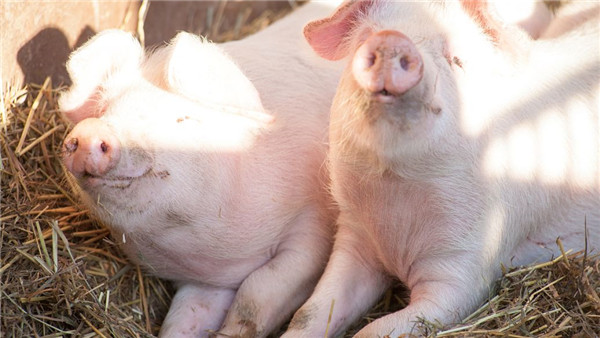 “非瘟”为何如此肆虐横行?猪场如
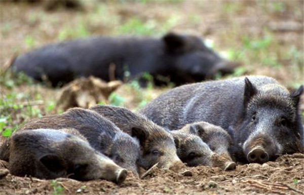 猪慢性传染病圆环病毒病的知识点
 脚下有泥,心中有光 | 牧原农艺师
 携手共赢,友谊长存:江西正邦作物保
 国新办举行7月份国民经济运行情况新
 农业农村部就2022年“三夏”生产形势